Fluorescence Microscopy (FM)
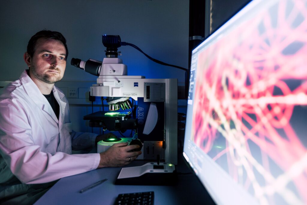
Zeiss Imager M2.m / Zeiss
Clear, focused images of sample regions enhance the analysis of biological markers or microfluidic chip structures. The Axio Imager M2's advanced features, such as the contrast manager and light manager, ensure defined conditions and reproducible results, facilitating precise and reliable observations.
UV light excitation fluorescence samples can be studied in detail. For some materials, intrinsic fluorescence might be present (e.g. dyes for light emitting diodes) while for other samples selective marking / staining can be used to study the presence and distribution of components (e.g. biomarker, uptake of nanoparticles in cells). Various additional light contrast methods, including brightfield, darkfield, and differential interference contrast (DIC), provide comprehensive imaging capabilities. Z-scan imaging allows for a 2.5D sample reconstruction.
A microfluidic device hosts areas that allow to adsorb selectively DNA of a virus. On development, the DNA is fluoresence labeled. To identify the amount of DNA as well as its distribution on the microfluidic chip, fluorecence microscopy is used. From the results an optimized SOP as well as an optimized chip geometry can be derived.